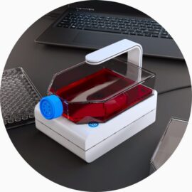
Live-cell imaging is a useful tool for monitoring the quality of cell cultures. A simple but effective set-up utilizing a high quality camera allows researchers to evaluate cellular health at regular points in time. The CytoSMART Lux2 duo-kit is a compact and cost-effective method for carrying out this type of analysis, following the complete growth progression of mammalian cells.
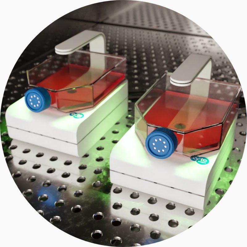
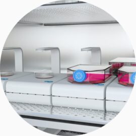
The CytoSMART Lux2 Duo Kit is an compact automated system, specifically designed to operate from within CO2– incubators and hypoxia chambers. Two devices operate via a single laptop, saving precious lab space. Gathering real-time insight into the progression of cell growth is now completely non-invasive. Bright-field imaging is deployed to create real time time-lapse videos, accessible remotely. Samples are also imaged under identical conditions, providing a robust platform for unambiguous comparison between cell culture variables, retaining data integrity.
Deploying this dual camera mini live cell imaging system is particularly useful in instances of stem cell research. The remote functionality of the CytoSMART cloud allows researchers to gain insight into the current confluence levels on their phone. This type of microscopy avoids over-growing of cultures or the loss of stemness in the cell-line, ensuring optimal yield. Your team can inspect the colonies that are formed by iPSCs and the morphology of MSCs without setting foot in the lab. Reducing the time spent in the lab will be crucial for many groups that try to continue research while complying to the social distancing rules
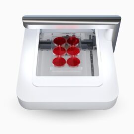
Inspect morphology and analyze cell confluency
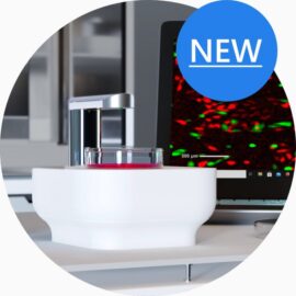
Compare cell cultures, supported by robust image analysis software
Imaging at regular time-intervals allows researchers to make quick decisions around cell quality, as any negative indicators such as blebbing, cell rounding, multi-nucleation and vacuole formation can be easily recognized. Variability in growth speed is easily recognized using the CytoSMART cell monitoring software, while image analysis provides an unambiguous read out for cell confluency meaning you can be confident in your data’s integrity. As such, the system is the ideal approach for the systematic comparison of cultures, in order to select the ideal culturing conditions.
Keep cells in a controlled environment
Complete cell culture incubator compatibility
During the observation of cell cultures, the right environmental conditions are vital in order to maintain cell health and keep cells active. Using a remote system avoids exposing the cells to potential mechanical and environmental shock from removing them from the incubator. Once the samples are placed inside the incubator, they no longer need to be handled. The researcher can check videos of the cell cultures in real-time, providing immediate insight into the culture quality. No need to step into the lab for routine inspection rounds, making the process efficient, cost effective and safe.
Technical specifications
| Optics | bright-field only with digital phase contrast |
|---|---|
| Magnification | 10X fixed objective – 20X digital zoom |
| Fluorescence filters | N/A |
| Camera | 5 MP CMOS |
| Data formats | JPG, XLSX, MP4 |
| Image size | 960 x 960 pixels |
| Field of view | 1.84 x 1.84 mm |
| Culture | well-plates, petri dishes, flasks, microfluidic chips and custom culture vessels |
| Computer requirements | Windows 10 – 2 USB3 ports |
| Power supply | AC 100-240V, 2A, 10W, 50/60HZ |
| Dimensions | 133 x 90 x 100 mm (L x W x H) |
| 5.2 x 3.5 x 3.9 in (L x W x H) | |
| Weight | 0.5 kg (1.1 lb) |
| Operating conditions | 0 – 42 °C, 5-95% humidity |
| Warranty | 1-year parts and labor |
| Data storage | unlimited cloud storage |
| Research use only. Not intended for diagnostic purposes. |


